
 PT
PT EN
EN ES
ES FR
FR DE
DE IT
IT HI
HI AR
AR ZH
ZH UZ
UZ TR
TR RU
RU ID
ID VI
VISpeak your findings.
Select the exam template. Dictate the anatomy, measurements and relevant changes in your own words.
Your attention belongs in the exam room. Dictate your findings and let Sono Ai Report help turn them into a structured report, ready for your clinical review.
The same demo as the main landing: total abdomen preset, original AI-template cards and both recording modes. The demo interface follows the page language; the example dictation is in Portuguese.
Open voice test in its own window ↗From a straightforward exam to a report that needs careful measurements and structured classifications. Keep the workflow together.
Select the exam template. Dictate the anatomy, measurements and relevant changes in your own words.
Transcription and report generation are distinct steps. The platform organizes your input around the selected exam model.
Check the findings, measurements and conclusion. Edit what is needed before signing and delivering your report.
Finish your dictation and generate a draft for your review, using the selected exam template.
Inspect and correct the transcript, then request report generation when you are ready.
You dictate or type the findings. The platform structures them, standardizes measurements and applies your templates, phrases and rules.
Total abdomen. Liver with mildly increased echogenicity, grade one steatosis. Simple cyst in segment seven, 1.8 cm. Gallbladder with a single mobile stone, 1.2 cm, with posterior shadowing. Right kidney with a simple cortical cyst, 2.1. Everything else within normal limits.
Liver: normal dimensions and contours. Homogeneous parenchyma with mildly increased echogenicity — grade I hepatic steatosis. Highlighting:
Simple anechoic cyst in segment VII, measuring 1.8 cm.
Bile ducts: intra- and extra-hepatic ducts of normal caliber.
Gallbladder: normal walls, containing a mobile stone with posterior acoustic shadowing, 1.2 cm.
Pancreas: normal shape and echotexture, no focal lesions.
Spleen: normal dimensions, homogeneous echotexture.
Right kidney: normal dimensions, preserved corticomedullary differentiation. Highlighting:
Simple cortical cyst measuring 2.1 cm.
Left kidney: normal, without calculi, cysts or pyelocaliceal dilatation.
Urinary bladder: adequately distended, thin walls, no lesions.
Use a lapel microphone and a configured presenter remote to capture exams in sequence. Return to your computer to review the individual transcripts and generate the reports.
A computer with internet access, compatible audio equipment and the recording window correctly configured. No phone required for this setup.
Watch the complete method: setup, dictation, transcript review and final report.
Watch the video · 4:50 ↓Scroll through the 15 original storyboard scenes. Select an image to enlarge it. Skip to the templates ↓
Clip the lapel microphone close to your mouth and plug its receiver into the computer's USB port. Before the first patient, press the ˅ button on the presenter once: the RECORDING light comes on and the level bars move as you speak. Press ˅ again to stop and delete that test card from the queue; it never turns into a report on its own.
In the Laudário, open the Sequential Recordings module: the window goes fullscreen by itself; if it does not, press F11. Stay in it for the whole shift: switching pages, closing the tab or letting the computer sleep closes the microphone, and the recording in progress is lost.
Keep the presenter in the chest pocket of your coat, where your hand finds it without looking. It comes pre-mapped: ˅ starts and stops the recording; VOL + starts another exam for the same patient. To change any button, use the presenter chip at the top of the window and press the key you want.
Press ˅ and say, before anything else, the patient's name, age and the exam: 'Maria Silva, 42 years old, total abdomen'. That is how the card is born filled in. If the name is not spoken, the card stays 'No name yet' and you type it at review; if the exam is ambiguous, pick it on the card with 'Choose exam'. Nothing is invented.
If you misspeak the patient's name or the exam type, cancel the dictation by saying 'cancelar gravação' (the command is recognised in Portuguese) and press ˅ to stop. The card is born red in the queue, never becomes a report and does not hold up the batch; start the same patient again with one click on the presenter. False alarm? Use 'Not a cancellation — restore exam' on the card.
Introduce yourself, explain what you are going to do and keep the conversation natural: the microphone stays open and does not need to be paused. Talk to the patient in your usual tone and keep the dictation for the findings; at review you confirm that the conversation stayed out of the report.
Position the patient, apply the gel and adjust the equipment while the recording runs. Silence costs nothing and does not cut the recording; do not press ˅ to 'pause', because stopping closes the card and the next press opens a new one.
Dictate in the order you scan: structure, finding, measurement and unit, spoken in words, 'gallbladder with a stone of eight millimetres'. Always state the side, right or left, and the unit for every measurement; the report only carries what you said.
Keep guiding the patient during the exam: 'breathe in, hold, now relax'. Speak instructions in one tone and findings in another, so that at review it is easy to confirm that the conversation was separated from the clinical findings.
If you said something wrong, say 'correcting' and repeat the whole sentence the right way: 'correcting, the correct measurement is twelve millimetres'. Do not just repeat the number. At review, check that the transcript kept the corrected value.
Finish the exam, agree with the patient how and when the report will be delivered and say goodbye with the microphone still open; none of this goes into the report. Only the next step, ˅, closes this recording.
Press ˅ once more: the RECORDING light goes out, the card appears in the queue with the patient's name and the audio goes to transcription on its own. Wait until 'Saving the recording…' disappears before calling the next patient; while it transcribes, the card shows 'Transcribing'.
Next patient: press ˅ and say the new name, age and exam. Another exam for the same patient: press VOL + instead; the card inherits only the name and age from the previous one, and you choose the exam later. While recording, VOL + is ignored, so pressing it by accident breaks nothing.
Back at the computer, open each card and read the transcript: name, age, exam, findings, measurements and side. Play the recording from the card if in doubt, correct the text in the card itself, pick the exam if it says 'Pick the exam', then mark 'Reviewed'. Only reviewed cards enter the batch; the queue never generates a report on its own.
At the end of the shift, press 'Generate N reports' once: the batch is generated in recording order and each card moves to the Completed tab with the 'Report ready' button. Open each report, make the final medical review, edit what is needed and only then finish and sign it.
Open a template and explore the real platform controls. These demos use example reports, with copying and export restricted.
Mark vessel paths, diameters and reflux on the diagram. Watch the platform's visual workflow in this real screen recording.
Reference tools support clinical judgment; they do not replace it. Check each tool's sources, scope and limitations.
Attach what is worth keeping: interesting cases, images, tables and documents that help you write the report. Every item starts private — you decide what stays with you and what goes to the community.
Quick reference and worked examples, with the source of every table. References support clinical judgement; they do not replace it.
Ultrasound, CT, MRI, X-ray and mammography share the same account and subscription environment.
Explore the same real method modules as the main landing. Mammography uses manual templates; the other methods use modular templates. Copying and export are disabled. Demonstration interfaces are currently in Portuguese.

Sono Ai Report was created by Dr. Pedro Pontes. UltrasoundReport.ai is its international gateway: the same platform, with a focus on ultrasound reporting.
CRM 14742 · RQE 9622 · Brazil
No. This is the international introduction to Sono Ai Report. Every platform-access button takes you directly to the official sign-in page at sonoaireport.com.
Yes. You can use manual reporting, structured modular templates or AI-assisted text. Choose the workflow that suits the exam.
The platform runs in your browser. The sequential-recording setup uses a computer, a compatible lapel microphone and a configured presenter remote.
No. AI output is a draft for professional review. Verify the transcription, findings, measurements and conclusion before signing or delivering any report.
No. Unlimited reporting refers to the manual and modular modes. AI features use the allowance or credits associated with your plan. Current trial and subscription conditions are shown in the platform.
Explore Sono Ai Report and find the reporting mode that fits your clinical routine.
Go to Sono Ai ReportOfficial sign-in · Sono Ai Report account · Physician review required
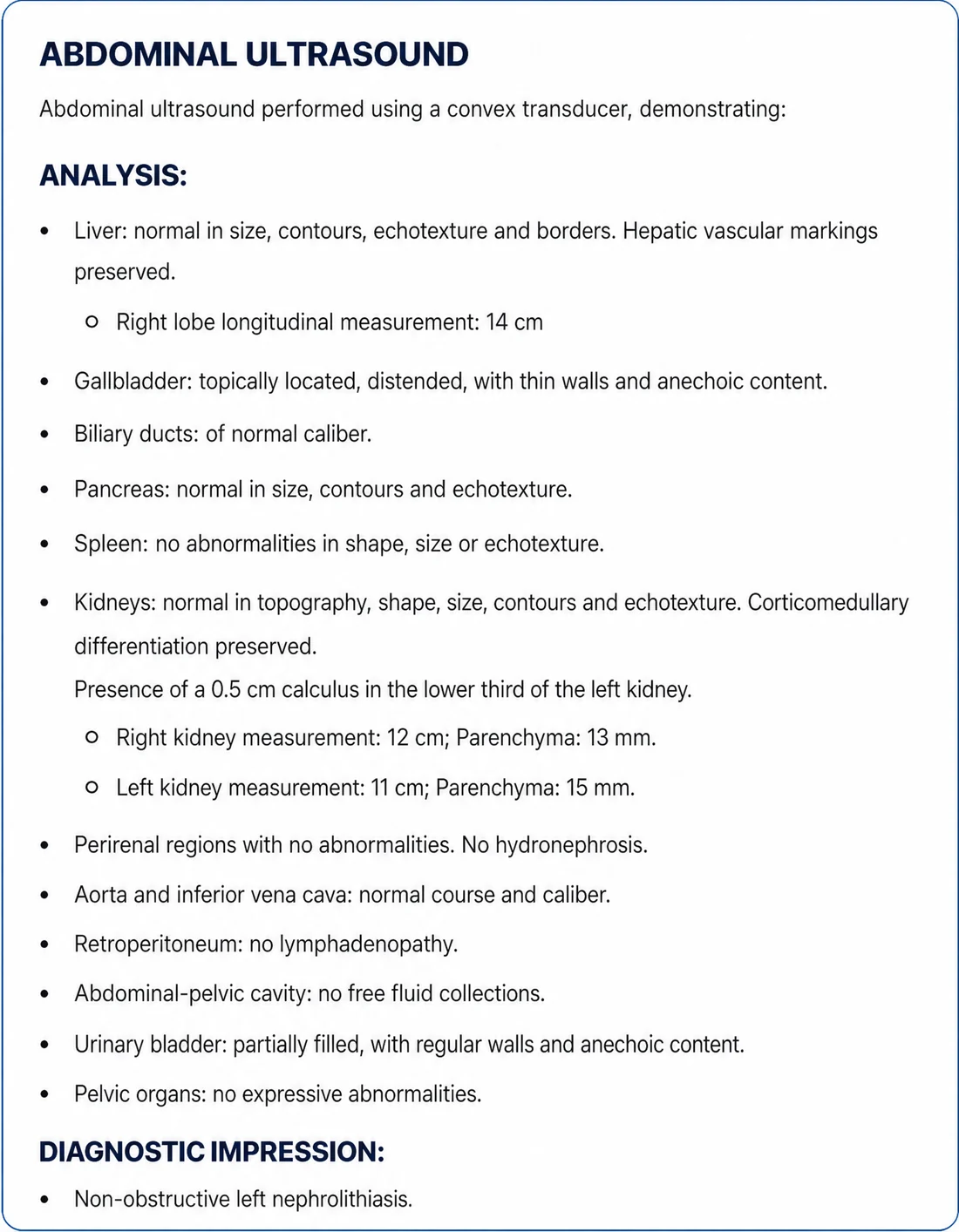
The report comes ready to review and sign — and you deliver it however you prefer.
Two fronts under construction. Neither is live today. When they ship, they are included in every plan, at no extra cost for anyone who already subscribes.
7-day free trial with 10 AI credits. Manual and modular reports are unlimited on every plan — and never use credits.
US$4.49/month
Unlimited
Uses AI credits
US$14/month
Unlimited
Uses AI credits
US$29/month
Unlimited
Uses AI credits
US$59/month
Unlimited
Uses AI credits
US$99/month
Unlimited
Uses AI credits
US$199/month
Unlimited
Uses AI credits
Monthly prices in US dollars (USD). AI credits cover voice dictation, text-assisted reports and AI specialists; manual and modular reports never use credits. Every report is reviewed and signed by the physician.